비뇨기과 검사방법
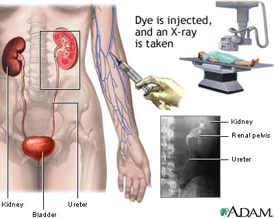
IVP
VCUG
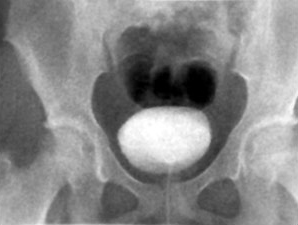
foley 카테터 같은 것으로 방광을 contrast 물질로 채운후 소변을 보게하면서
fluoroscopy(연속적인 촬영)로 본다
Retrograde pyelography
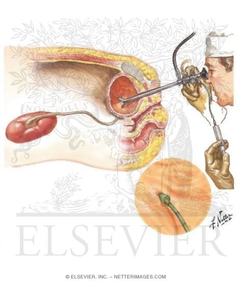
Retrograde pyelography combines the use of a long, flexible
viewing tube called a cystoscope with contrast x-rays to visualize the kidneys
and ureters. The cystoscope is inserted through the urethra into the bladder;
fiberoptic cables permit direct visual inspection of these structures. A
catheter is then threaded through the scope so that a contrast dye can be
infused directly into the ureters to delineate them on x-ray films. Retrograde
pyelography is most often performed when intravenous pyelography produces
inconclusive results, or when it cannot be performed because of impaired kidney
function or another reason.
방광경으로 보면서 다이를
ureter에 맞춰서 쏜다. 그러면서
fluoroscopy로 촬영한다
신기능이 나빠서 IVP하기 어려울때, IVP검사 결과가 애매할때
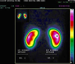
DMSA(APN의 진단에 매우 sensitive)
radionuclide
emits a type of radioactivity called gamma ray
injection into a vein
DMSA is used because it builds up concentrate in the kidneys
areas of the target organ or tissue which emit lots of gamma rays may be shown as red spots ('hot spots')
목적 : to check the structure of the kidneys, their size and shape
children who have had urinary tract infections
areas of the kidney are working well and any areas of scarring
Scarring can be caused by a condition in which urine travels back from the bladder to the kidneys. This is called vesico-ureteric reflux
also look for reduced blood supply to the kidneys.
DMSA does not attach itself to areas of the kidneys that are damaged.
monitor any changes to inflammation of the kidneys.